Diagnostic
Diagnostic Medical Sonography (DMS), referred to as ultrasound, is a medical diagnostic procedure that uses high-frequency sound waves to produce dynamic visual images of organs, tissues, or blood flow inside the body. The procedure uses sound waves and their echoes to form two or three-dimensional images. These sound waves travel into your body and are reflected by different tissues and organs back to the ultrasound probe.
An ultrasound may be performed to detect or diagnose problems with the:
- Abdomen: Detects abnormalities in the kidneys, liver, pancreas, and gallbladder.
- Breast: Examines palpable masses in breast tissue or characterizes masses seen on a mammogram.
- Carotid: Shows where blood flow is restricted on its way to the brain or kidneys.
- Doppler: Examines the body’s interior structures while simultaneously evaluation the flow of blood.
- Interventional: Helps Radiologists see precisely what they are doing while performing aspirations, biopsies, injections, or other procedures.
- Musculoskeletal: Examines pain in joints or muscles and looks for problems such as muscle or tendon tears, abnormal fluid collection, or masses.
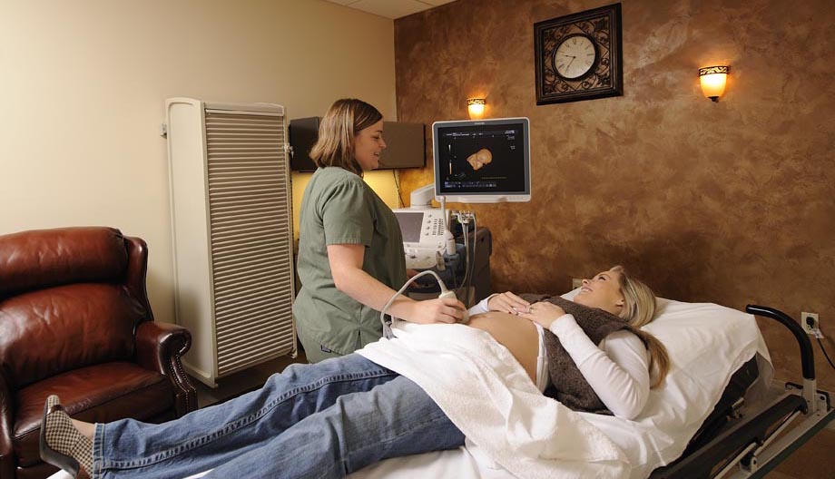
- Obstetric: Monitors fetal development.
- Pelvis: Assesses bladder, kidneys, ovaries, and uterus in an attempt to locate reasons for pelvic pain.
- Retroperitoneal: Studies the kidneys and urinary tract.
- Scrotum: Attempts to locate reasons for testicular pain, swelling, and/or lumps.
- Thyroid: Examines the thyroid gland and the region for anything suspicious or abnormal.
- Vascular: Examines the vascular system and detects blood clots and narrowed arteries.
Therapeutic
Therapeutic sonography is used for detection and aspiration of fluids in various cavities of the body. These procedures are more likely to give relief to patients. Some of these procedures include aspiration of fluid from the abdominal cavity (paracentesis), plural effusion (thoracentesis), or guidance when an intravenous line is required but the vein is difficult to locate.
What to Expect
The patient may be lying in different positions depending on the exam and/or circumstance. A warm non-allergenic gel is applied to the skin for sound conductivity. The sonographer moves the transducer (probe) against the skin and sweeps it back and forth over the area being examined. If the area the sonographer is scanning is sensitive or tender, the patient may feel some discomfort or pain. Once the exam is complete, the gel is wiped off the patient’s skin. The sonographer will consult with the radiologist and the patient will be released to resume his/her normal activities unless indicated otherwise.
How to Prepare
Most of the exams do not require preparation.
Abdominal ultrasounds require that you don’t eat, drink, or smoke for approximately 8 hours prior to the exam.
Pelvic and Obstetric ultrasounds require that you drink 32 ounces of water to fill your bladder 2 hours prior to the exam.
Mountain View Hospital Imaging Center | Phone: 208-557-2887 | Fax: 208-557-2746